Part of the The Complete Guide to Spiders: Identification, Prevention & Removal guide.
A spider is built entirely around the act of hunting. Every major anatomical system, the eyes, the legs, the chelicerae, the silk glands, the venom apparatus, is a tool shaped by hundreds of millions of years of predatory specialization. Understanding how these structures work explains why spiders are so effective at what they do, and why so many features that seem strange at first make perfect mechanical sense on closer examination.
For a comprehensive overview, see our Complete Guide to Spiders.
Two Body Segments
All spiders have exactly two body segments. This immediately distinguishes them from insects, which have three, and from most other arthropods. The two segments are the cephalothorax and the abdomen, connected by a narrow, flexible stalk called the pedicel.
The pedicel is more than a simple joint. It allows the abdomen to rotate and move independently of the cephalothorax, enabling a spider to point its spinnerets in any direction while keeping all eight legs firmly planted on a surface or web. Without pedicel flexibility, the precision of silk manipulation that defines spider web-building would be impossible.
The Cephalothorax
The cephalothorax (also called the prosoma) is the anterior segment, a fused head and thorax covered on top by a hardened plate called the carapace. It contains or supports every structure the spider uses to locate, pursue, and subdue prey:
- The brain and central nervous system
- The eyes (positioned on the front and sides of the carapace)
- The chelicerae (fangs) and associated venom glands
- The pedipalps (sensory and, in males, reproductive appendages)
- Attachment points for all eight legs
- The sucking stomach
A visible groove or pit in the center of the carapace, called the fovea, marks where powerful internal muscles attach to the sucking stomach. The fovea's position and shape are useful features for family-level identification in many spider groups.
The Abdomen
The abdomen (opisthosoma) is the posterior segment and is almost entirely soft-walled, making it the spider's most vulnerable body part. It houses:
- The heart and open circulatory system
- Digestive glands and the mid-gut
- Reproductive organs
- Book lungs and tracheal tubes (respiratory systems)
- All major silk glands
- The spinnerets
Eyes: Number, Arrangement, and Function
Most spiders have eight eyes, but the number varies by family. Some have six, four, two, or even no functional eyes at all. Eye arrangement, the specific pattern of eye positions on the face, is one of the most reliable characters for spider family identification and is used extensively in taxonomic keys.
All spider eyes are simple ocelli (single-lens structures), not compound eyes like those of insects or crustaceans. They divide into two functional categories:
- Principal eyes (anterior median eyes, AME): Forward-facing, typically the largest eyes, capable of high spatial resolution. In jumping spiders, these are elongated tube-shaped structures with a movable retina that the spider can scan independently to track targets without turning its head.
- Secondary eyes (all remaining eye pairs): Arranged around the carapace to cover a wide field of view, these detect motion and changes in light intensity. Many secondary eyes contain a tapetum lucidum, a reflective layer that produces the characteristic eyeshine visible when a wolf spider is illuminated with a headlamp at night.
| Spider Family | Eye Count | Arrangement | Key Visual Feature |
|---|---|---|---|
| Jumping spiders (Salticidae) | 8 | AME large, forward-facing | High-resolution color vision, movable retina |
| Wolf spiders (Lycosidae) | 8 | Three rows | Strong tapetum, motion detection |
| Orb-weavers (Araneidae) | 8 | Two rows of four | Moderate resolution |
| Brown recluse (Sicariidae) | 6 | Three pairs in curved row | Limited, adapted for dim conditions |
| Cave-dwelling species | 0 to 2 | Vestigial or absent | Absent, compensated by mechanoreception |
Chelicerae and the Venom System
The chelicerae are the paired appendages at the front of the cephalothorax that terminate in the fangs. In araneomorph spiders (the majority of North American species), the chelicerae fold inward, closing toward each other in a pinching motion like a pair of scissors. In mygalomorph spiders (tarantulas, trapdoor spiders, funnel-webs), the chelicerae are oriented to strike downward like parallel pick-axes.
Each chelicera contains a hollow fang connected by a duct to a venom gland sitting within the cephalothorax. When the spider bites, muscles compress the venom gland, forcing venom through the duct and out a small opening near the fang tip. The spider can regulate the volume of venom injected and frequently delivers "dry bites" with little or no venom when biting defensively, reserving venom for actual prey capture.
According to the CDC, most spider bites produce minimal medical effects in part because defensive bites commonly involve partial or no venom injection. The spider's evolutionary priority is prey capture, not human defense.
Legs: Eight-Legged Engineering
Spiders have eight legs, each with seven distinct segments: coxa, trochanter, femur, patella, tibia, metatarsus, and tarsus. The tarsus ends in two or three claws used to grip surfaces and manipulate silk threads. A structure called the claw tuft, dense adhesive setae between the claws, allows spiders to walk on smooth vertical surfaces by molecular adhesion rather than mechanical gripping.
Leg movement in spiders is controlled by a combination of muscular flexion and hydraulic extension. Spiders can only flex their legs using muscles; extension requires hemolymph pressure pushing outward from within. This hydraulic dependence explains why dead spiders always curl inward: without circulatory pressure, the flexor muscles have no antagonist and pull the legs toward the body by default.
Each leg is also a sophisticated sensory array. Slit sensilla (fine mechanoreceptors arranged in groups on leg surfaces) detect substrate vibrations, air currents, and mechanical deformation with extraordinary sensitivity. Chemoreceptor hairs on the tarsi allow spiders to taste surfaces by contact, effectively sampling the chemical environment with every step.
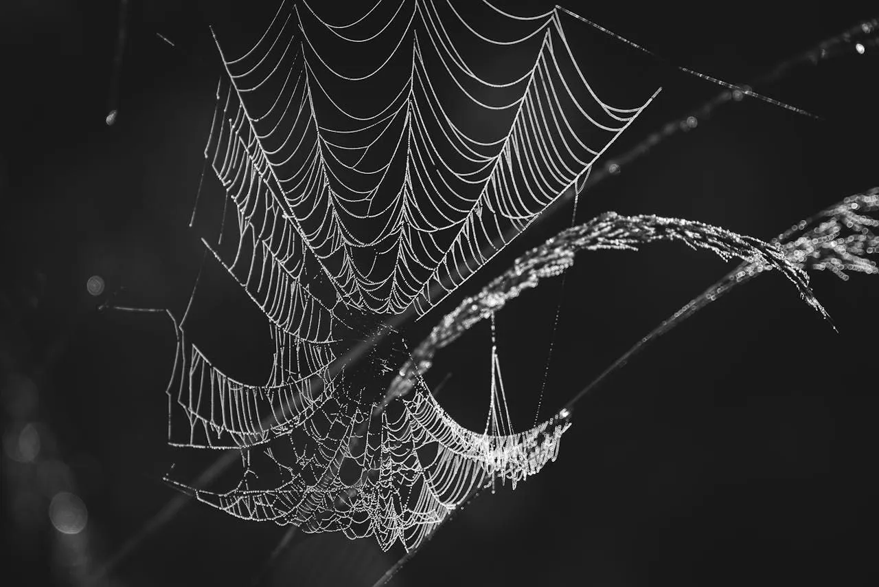
Silk Glands and Spinnerets
Spider silk is produced in specialized abdominal glands and extruded through spinnerets, finger-like appendages at the posterior of the abdomen. Most spiders have three pairs of spinnerets (six total), each connected to different gland types producing different silk chemistries.
Major silk types and their functions:
- Major ampullate silk (dragline): The structural silk used for web frames, radii, and the spider's personal safety line. Gram for gram, one of the strongest biological materials known, with tensile strength comparable to high-tensile steel and extensibility far exceeding it.
- Viscid or cribellate silk (capture thread): Sticky or woolly silk deployed across the spiral of orb webs to intercept and hold prey.
- Tubuliform silk (cylindrical): Thick, tough silk used exclusively for constructing egg sacs.
- Piriform silk (attachment cement): The silk used to bond other silk to surfaces, essentially the spider's structural adhesive.
According to the USDA, research into synthetic spider silk has been ongoing for decades because the combination of strength, flexibility, and biocompatibility has potential applications in textiles, sutures, and advanced materials. No synthetic process has yet matched the properties of natural spider silk produced at ambient temperature and pressure.
Respiratory and Circulatory Systems
Spiders use two types of respiratory structures, sometimes in combination. Book lungs are leaf-like organs in the anterior abdomen where hemolymph flows between thin lamellae (pages) exposed to an air space, exchanging gases passively. Tracheae are tubes that carry air directly to tissues, reducing reliance on circulatory transport for oxygen delivery.
Mygalomorphs (tarantulas and their relatives) rely primarily on two pairs of book lungs. Many araneomorphs retain one pair of book lungs and supplement them with a tracheal system, and in very small spider species, book lungs are often reduced to vestigial structures with tracheae taking over almost completely.
The circulatory system is open: hemolymph (spider blood, containing the blue oxygen-carrier hemocyanin) circulates through body cavities rather than enclosed vessels. The heart is a simple muscular tube running along the dorsal abdomen, pumping hemolymph forward into the cephalothorax and out through the body cavity.
In my 15 years in pest management, spider anatomy is the topic I've found most effective for changing client attitudes. Once someone understands that spiders can regulate venom delivery and that most defensive bites involve minimal venom, the entire risk conversation shifts. Anatomy turns fear into curiosity, which is exactly where productive pest management thinking begins.
How to Identify
Spider anatomy provides the most reliable identification tools. Eye count and arrangement are family-level identification characters: six eyes in three dyads identifies the Sicariidae family (brown recluse and relatives); two large forward-facing eyes flanked by smaller rows identifies jumping spiders (Salticidae); large top-head eyes in a wolf spider (Lycosidae). Chelicera orientation separates two major groups without a microscope: araneomorphs (most North American species) have chelicerae that fold inward, closing side-to-side; mygalomorphs (tarantulas, trapdoor spiders) have chelicerae that strike downward. Body proportions aid identification: bulbous, rounded abdomens on cobweb weavers; elongated, tubular abdomens on cellar spiders and some orb weavers; compact bodies on jumping spiders and crab spiders. These anatomical markers allow confident identification without handling the spider.
Risk and Severity
Anatomical features predict medical risk more accurately than color or size. Fang orientation matters clinically: mygalomorph fangs strike downward and are typically large enough to penetrate skin reliably but produce less medically significant venom in North American species. The presence of venom glands is nearly universal in spiders, but the composition varies enormously - most species produce venom active only against insect prey with negligible effect on humans. Per the CDC, medically significant spider bites in North America come almost exclusively from two groups: Latrodectus (black widows), identifiable by the hourglass marking, and Loxosceles (brown recluses), identifiable by six eyes in three pairs and the violin marking. Anatomical verification before assuming medical risk prevents both panic about harmless species and complacency about dangerous ones.
Solutions and Actions
Apply anatomical knowledge before committing to any control action. Confirm the family before treating: a spider with eight eyes cannot be a brown recluse - no matter how similar it looks - and does not warrant recluse-level intervention. Use the chelicera orientation to confirm mygalomorph (tarantula, trapdoor spider) versus araneomorph. For medically significant species, document with a macro photograph capturing the eye arrangement and submit to your local cooperative extension for confirmation before applying chemical treatment. Physical removal with a jar and card is appropriate for all non-venomous species; professional removal is appropriate for confirmed widow or recluse populations in living spaces.
Prevention
Anatomical knowledge guides prevention priorities. Understanding that spiders use adhesive setae on their tarsi to climb smooth surfaces explains why smooth paint, glass, and metal surfaces are less likely spider pathways than rough brick and textured siding - concentrate exclusion efforts at gaps in smooth-surface areas where spiders cannot simply climb past a partial seal. Knowing that spiders walk on leg tips rather than dragging their bodies explains why diatomaceous earth applied at leg-joint contact points is more effective than broadly applied liquid residuals. Understanding silk gland anatomy explains why egg sac removal is a high-priority intervention: each egg sac represents hundreds of offspring that the spider invested significant silk resources to produce and protect.
Main Causes
Indoor spiders activity reflects two drivers — a hospitable indoor environment and a sufficient supply of insect prey. Spiders enter through gaps under doors, around windows, utility penetrations, and any opening leading to attics, basements, garages, or crawl spaces. Once inside they settle wherever undisturbed corners, low light, and easy prey access converge. Cooler weather pushes outdoor species inside in late summer and fall as they seek mating sites or shelter. The most important upstream driver is the indoor insect population — homes with active fly, gnat, moth, or other pest activity sustain larger spider populations than homes without prey. Cluttered storage areas, accumulated webbing, and outdoor lighting that draws nocturnal insects all amplify the indoor pressure.
Frequently Asked Questions
Why do dead spiders curl up?
Dead spiders curl inward because leg extension requires active hydraulic pressure from circulating hemolymph. When circulation stops at death, pressure drops and the only forces acting on the legs are the flexor muscles, which pull inward. The result is the characteristic curled posture visible in any dead spider.
Do spiders have brains?
Yes. Spiders have a central nervous system concentrated in a ganglionic mass within the cephalothorax. In some smaller spider species, the brain is proportionally so large it extends physically into the legs, a literal anatomical reality documented by researchers at the Smithsonian Tropical Research Institute studying tiny tropical species.
What is the pedicel and why does it matter?
The pedicel is the narrow, flexible stalk connecting the cephalothorax to the abdomen. It allows the abdomen to rotate and angle independently of the front body while the spider's legs remain stable on a surface. This flexibility is essential for precise spinnneret positioning during web construction, egg sac building, and silk attachment. It is one of the features most responsible for the astonishing range of silk structures spiders produce.
What should I recheck first for spider anatomy?
Recheck the exact place, timing, and repeated signs connected with spider anatomy before changing your plan. A single sighting or old web can mean something very different from fresh activity in several rooms. Confirm whether insects, clutter, moisture, gaps, or stored items are supporting the issue, then match the response to what you actually found.
Continue reading:
The Complete Guide to Spiders: Identification, Prevention & Removal →Sources & Further Reading
- Venomous Spiders — U.S. National Institute for Occupational Safety and Health
- Spiders — Pest Notes — University of California Statewide IPM Program
- Insect Stings and Bites — American Academy of Allergy, Asthma & Immunology